Equine-Thorax : A case of viral bronchopneumonia in a horse
French Warmblood 20 years old. presented for hyperthermia and anorexia since a week.
A mild expiratory dyspnea is noted; the lung sounds are modified, crackles and wheezes are detected.
Normal cardiac auscultation.
A mild expiratory dyspnea is noted; the lung sounds are modified, crackles and wheezes are detected.
Normal cardiac auscultation.
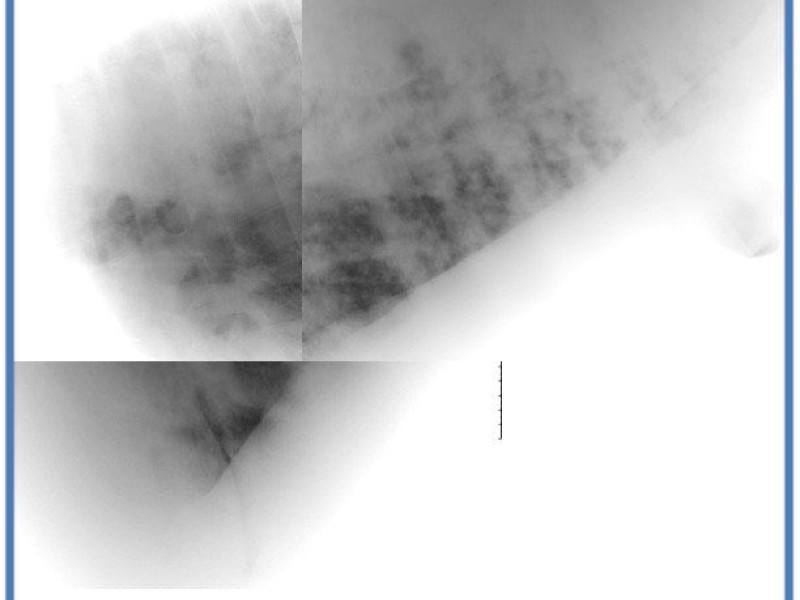
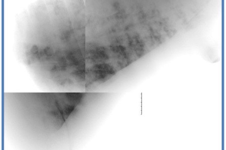
A patchy, cloudy alveolar pattern is detected throughout the lung field, more prominent in the caudodorsal lung lobes. The alveolar patches show fluffy margins. A diffuse peribronchial pattern is also detected and give an impression of coalescing nodules at some point.
The differentiel diagnosis include
- a bronchopneumonia or infectious or parasitic origin.
- a neoplasia with metastatic vs primary diffuse bronchogenic carcinoma or squamous cell carcinoma.
Exercice induced pulmonary hemmorhage or pulmonary edema are other differentials but not likley in this case considerng the age and activity of the horse.
A BAL and pulmonary biopsies have revealed a diffuse bronchopneumonia secondary to equine herpes virus type 5.