Canine-Thorax : A case of non cardiogenic pulmonary edema secondary to heat shock in a french Bouledogue
5-year-old male French Bouledogue presented for severe acute dyspnea. The mucosal membranes of this animal are cyanotic. The dyspnea appeared after a road trip of several hours while coming back from holidays.
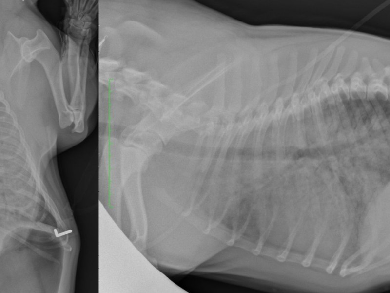
First of all we observe a large amount of gas within the stomach, which is a sign of severe respiratory distress (aerophagia).
Generalized peribronchial opacification of the lung field (thick donuts), and presence of few air bronchogram as a sign of alveolar pattern in some areas.
The coalescence of peribronchial infiltrates creates an impression of nodular or miliary opacifcation. The presence of gas within these small "nodules" rules out the presence of nodules and confirms the peribronchial infiltration.
The cardiac silhouette demonstrates a normal size and shape.
The vertebral bodies of T7 and T8 present a wedge and triangular shape (common pathology in brachycephalic dogs, commonly non clinically relevant).
Conclusion: radiographic signs compatible with a non cardiogenic edema or acute respiratory distress syndrom, most likely due to "heat" in the car. The animal received as an emergency treatment oxygen, infusion. Its body temperature was decreased thanks to the use of alcoolized swabs placed on its extremities. It did not receive any diuretics. The administration of diuretics, although very discussed and controversed in the literature, is not mandatory in non cardiogenic edema. A control chest xrays was performed and did not show any abnormality anymore.