Canine-Thorax : A case of oesophageal diverticulum secondary to the persistance of the right aortic arch
Labrador puppy of weeks.
Regurgitations since weaning.
Clinical exam and ausculation are normal.
Regurgitations since weaning.
Clinical exam and ausculation are normal.
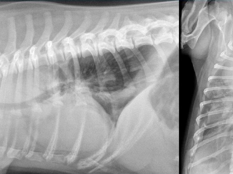
A gas filled element is visible overlying the thoracic trachea. There is cranial mediastinal widening on the VD and the gas filled structure is also present corresponding to the esophagus.
The trachea is deviated to the left focally on the VD and ventrally on the lateral view. The lumen is narrowed very discreetly.
Cardiovascular structures are normal. The lungs are normal. The thymus is visible on the VD view.
Conclusion:
Esophageal diverticulum and tracheal deviation on the left and ventrally.
This is pathognomonic of a persistent right aortic arch.
Esophageal stricture is created by the persistence of arterial ligament that connects the aorta and the left pulmonary artery. This ligament passes dorsally to the esophagus and creates an area of stricture.
recommendation:
A CT scan is recommended to check the anatomy of the vessels. In the case of this patient, malposition of subclavian arteries is present. The left subclavian artery also passes dorsally to the esophagus and is involved in the stricture.
Surgery was performed and involves cutting the arterial ligament.
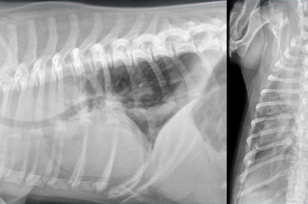