Canine-Thorax : A case of oesophageal diverticulum secondary to the presence of a right aortic arch in a young dog
English cocker, 3 months.
History:
Regurgitation since weaning
Stunted growth
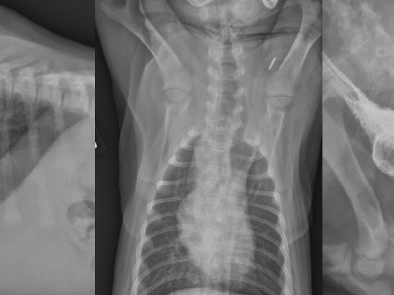
Radiography: thorax RL view native, RL view after positive esophagogram (barium).
History:
Regurgitation since weaning
Stunted growth
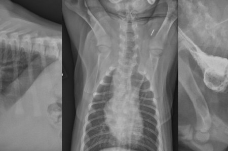
Radiography: thorax RL view native, RL view after positive esophagogram (barium).
Description:
Native radiographs: A round, well defined gaseous structure is visible in the cranial mediastinum. this strucure is extending in the neck through the thoracic inlet. The trachea is deviated ventrally in the thorax.
Esophagography: Contrast is seen in the previsously described structure which is the esophagus. There is a focal dilation of the esophagus.
The differential diagnosis is congenital diverticula, vascular ring anomaly, stricture and cranial dilation.
Such a focal dilation of the esophagus with regurgitation since weaning is compatible with a vascular ring anomaly and especially the persistance of the right aortic arch. The esophagus is comprised between the base of the heart, aorta, left ductus arteriosus and pulmonary trunk.
Surgery is necessary,