Canine-Thorax : A case of pneumomediastinum secondary to a tracheal rupture in a dog
Male Husky 1 year old, acute respiratory distress after a fight with another dog.
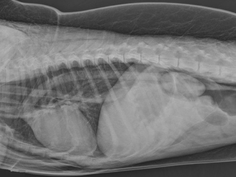
Radiography: RL thoracic radiograph.
Surprinsingly, both kidneys are very clearly visible, they are boredered by gas opacity. The intestinal mass is displaced ventrally by the gas dilated retroperitoneal space. This corresponds to a pneumoperitoneum.
There is also a severe subcutaneous emphysema visible.
In the cranial mediastinum, the cranial vena cava, the brachiocephalic trunk and the left subclavianartery are visible.
The dorsal border of the aorta is outlined by a gas opacity. These vessels are not normaly seen. This coresponds to a pneumomediastinum.
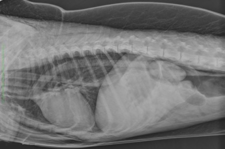
A small tear in the thoracic trachea was found on endoscopy.
Pneumomediastinum is secondary usually to a trauma with rupture of the trachea, main stem bronchi or esophagus or a wound in the soft tissues of the neck. A contusion of the lungs can also give rise to pneumomediastinum, with migration or air from the alveoli to the mediastinum.
Gas in the mediastinum can then follow the aorta in the aortic hiatus and migrate in the retroperitoneal space.