Canine-Abdomen : A case of paraprostatic cyst in a dog
9 year-old intact male American Staffordshire Bullterrier presented with abdominal distension, tenesmus and stranguria.
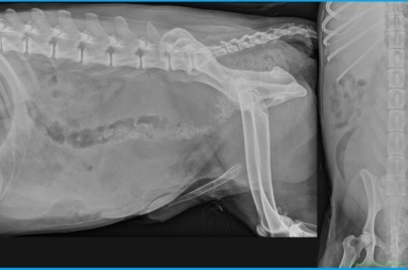
Abdominal radiographic examination.
Abdominal radiographic examination.
We can observe:
- the loss of serosal details in the caudal and mid abdomen;
- a marked ventral deviation of the descending colon and the presence of a large amount of fecal material within the rectum;
- absence of visualisation of the urinary bladder and prostate;
- presence of an ill-defined homogeneous soft tissue opacity within the caudoventral abdomen and of another mass with similar appearance in the dorsal aspect of the caudal abdomen;
- no radiopaque elements visible on the urinary tract;
- lombosarcal spondylosis.
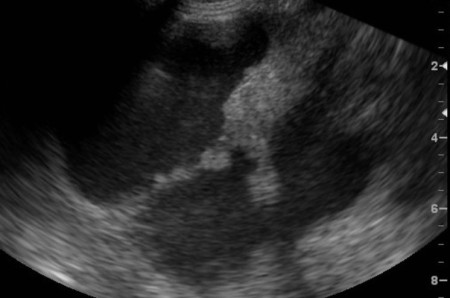
The differential diagnosis for caudal abdominal masses in an intact male dog presenting with tenesmus and stranguria is quite short: this can be due to a prostatic origin (paraprostatic cysts, prostatic abscess or tumour) or bladder origin although less probable given the size of the masses and the absence of periosteal reaction in the lumbar region (paraneoplastic syndrom). A paraprostatic cyst (or several cysts) is the major hypothesis. An abdominal ultrasound confirmed the radiographic diagnosis, and also to evaluate the number and size of the cysts, and eventually to guide aspiration of those cysts. A retrograde urogram might also allow the localization of the bladder.
- the loss of serosal details in the caudal and mid abdomen;
- a marked ventral deviation of the descending colon and the presence of a large amount of fecal material within the rectum;
- absence of visualisation of the urinary bladder and prostate;
- presence of an ill-defined homogeneous soft tissue opacity within the caudoventral abdomen and of another mass with similar appearance in the dorsal aspect of the caudal abdomen;
- no radiopaque elements visible on the urinary tract;
- lombosarcal spondylosis.
The differential diagnosis for caudal abdominal masses in an intact male dog presenting with tenesmus and stranguria is quite short: this can be due to a prostatic origin (paraprostatic cysts, prostatic abscess or tumour) or bladder origin although less probable given the size of the masses and the absence of periosteal reaction in the lumbar region (paraneoplastic syndrom). A paraprostatic cyst (or several cysts) is the major hypothesis. An abdominal ultrasound confirmed the radiographic diagnosis, and also to evaluate the number and size of the cysts, and eventually to guide aspiration of those cysts. A retrograde urogram might also allow the localization of the bladder.