Féline-Thorax : A case of peritoneopericardial diaphragmatic hernia in a cat
Male castrated cat, 6 years, apathic.
Auscultation: Muffled heart sounds, no abnormalities of the respiratory system detected.
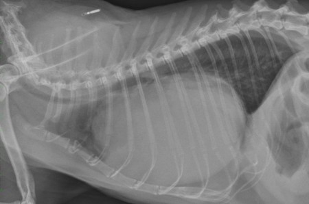
Radiography: lateral view of the thorax.
Auscultation: Muffled heart sounds, no abnormalities of the respiratory system detected.
Radiography: lateral view of the thorax.
Important remark: the liver is not visible in the cranial abdomen; the stomach and an intestinal loop (probably colon) are seen against the diaphragm.
The thoracic trachea is displaced dorsally by an enlarged cardiac silhouette. The cardiac silhouette occupies 90% of the thoracic height and 70% of the thoracic width. Its opacity is heterogeneous. The diaphragm is silhouetting with the cardiac shadow, a dorsal horizontal line is seen binding the heart shadow and the diaphragm (the pericardiodiaphragmatic mesothelial remnant).
The sternum shows a mild ventral curvature showing the chronicity of the disease.
Conclusion: pleuropericardiodiaphragmatic herniation (PPDH) with herniation of the liver and small intestinal loops.
This is a congenital anomaly. The Persian breed is predisposed and there is usually an association with a pectus excavatum.
It is often an incidental finding without clinical consequences, however when there is torsion or strangulation of one abdominal organ it can cause clinical signs. In this case on liver lobe was probably strangulated causing the apathy. The surgical treatment is recommended before 4 months, in order to avoid adhesions between the herniated organs.