Canine-Thorax : A case of pulmonary lobar torsion in a Pug
Pug, male, 2 years old.
Acute cough, episode of tachypnea since a few days. The owners report a collapse with cyanotic mucous membranes. Symptomatic treatment by the vet.
since then anorexia, mild lethargy and persisting cough.
On auscultation, the animal is quiet, respiratory rate is 42 per minute, heart rate 120. Pale mucous membranes, CRT normal. The respiratory sounds and heart sounds are muffled, more prominent in the left cranial thorax. Thoracic radiographs are taken.
Description:
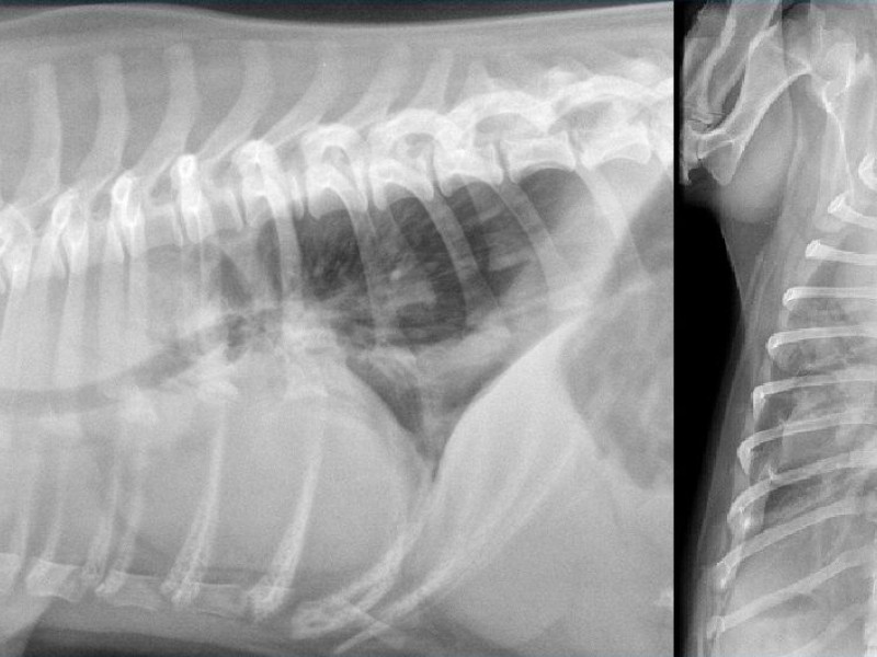
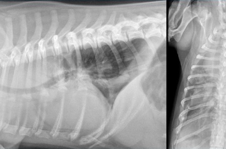
There is a marked dorsal deviation of the trachea cranial in the thorax. The cardiac silhouette is hardly discernible, it is masked by a soft tissue opacification type in the ventral thorax on the lateral radiograph. The costodiaphragmatic recesses are rounded and an interlobar fissure line is visible between the left cranial and caudal lobes. On the VD, only the left cranial aspect of the cardiac silhouette is obscured. It is continuous with a diffuse opacification of the left cranial lung lobe. If you look closely, you realize that this clouding is not homogeneous. We notice some radiolucent bubbles coalesced in the center of the lobe. On the lateral, some air bronchograms are visible, superimposed on the cranial aspect of the cardiac silhouette. We see an interruption of the left cranial bronchus few centimeters distal to the carina.
Conclusion:
Mild pleural effusion, and focused around the left cranial lobe. Left cranial lobe consolidation with a clouding which is specially called vesicular. Abrupt interuption of the left cranial bronchus under the carina.
Differential Diagnosis:
On a lobar consolidation, many differentials are possible like neoplasia, lobar pneumonia, thromboembolism, bleeding. A mediastinal mass can possibly be part of the list. In this case we have some interesting discoveries namely vesicular opacification of the lobe, focused effusion, an interruption of a bronchus. This is in favor of a lung lobe torsion.
Further examination:
Thoracic ultrasound is performed to visualize the lack of vascularization in the lobe. The venous signal is the first to disappear and then the arterial signal disappears. The lobe is typically hepatized, the fluid bronchograms are visible and sometimes air bubbles may be trapped in the middle of the lobe.
Treatment:
It is surgical in all cases, it is a lobectomy.
Discussion:
The twists of lobes may be spontaneous or secondary to a phenomenon such as pleural effusion, neoplasia, a trauma, a lobectomy. A partial collapse of the lobe changes the spatial relationships between the lobes and may cause its twist.
Large breed dogs such as Afghan hound are predisposed to spontaneous twists of the right medial lobe. In recent years, pugs appear as dogs prone to spontaneous twisting of the left cranial lobe.