Canine-Thorax : A case of hiatal hernia in a dog
English Bulldog, 2 years presented for chronic regurgitation.
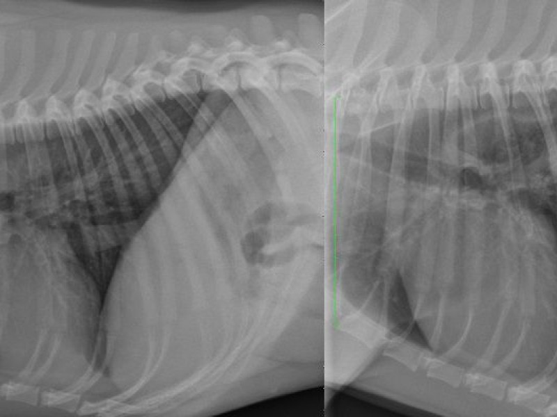
Thoracic radiographs (LLviews) with and without compression of the cranial abdomen with a radiolucent tool.
Thoracic radiographs (LLviews) with and without compression of the cranial abdomen with a radiolucent tool.
No radiographic anomalies are detected on the native radiographs. On the compression radiograph, a gas containing viscus is detected in the dorsocaudal lung field. Around this viscus, a second tubular viscus is detected. The stomach is not visible in the cranial abdomen.
These findings are consistent with a hiatal hernia. The compression views are usually necessary to see the herniation. A normal radiography without compression doesn't exclude a hiatal hernia. When fluoroscopy is not available this technique is interesting.
These findings are consistent with a hiatal hernia. The compression views are usually necessary to see the herniation. A normal radiography without compression doesn't exclude a hiatal hernia. When fluoroscopy is not available this technique is interesting.