Canine-Thorax : A case of pulmonary metastases of mammary gland carcinoma in a bitch
Female Shih Tzu, 10 years. Previous surgery of mammary tumors, 2 weeks ago. On histology, it appeared to be a high grade carcinoma with microembolism.
The dog is fine clinically.
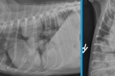
Radiography: RL and VD radiograph of the thorax.
The dog is fine clinically.
Radiography: RL and VD radiograph of the thorax.
Multiple nodules and masses of homogeneous soft tissue opacity and well-defined contours are seen in the lungs. Their shape and size are variable and their distribution randomized in the lung field.
This image of "canon ball' is typical for pulmonary metastases, particularly secondary to primary epithelial tumour.
This image of "canon ball' is typical for pulmonary metastases, particularly secondary to primary epithelial tumour.