Canine-Thorax : A case of mediastinal hemorrhage secondary to rhodenticides intoxication
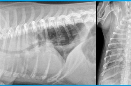
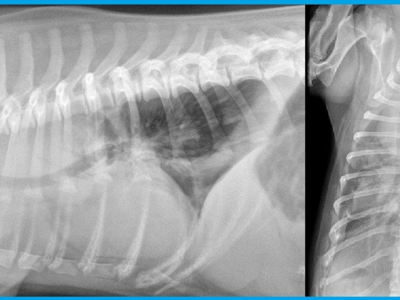
5-year-old Dachshund dog presented with acute respiratory distress, pale mucosa. Radiographic examinations: right lateral and dorsoventral projections.
Ventral deviation of the intrathoracic trachea. Presence of a soft tissue/liquid opacity in the cranial mediastinum in ventral and dorsal regions. The cranial mediastinum measures more than twice the width of the thoracic spine on the dorsoventral projection. Presence of fissure lines and liquid opacity between the rib cage and the lung borders; roundening of the lombo- and costo-diaphragmatic recesses. The tracheobronchic bifurcation is located at the 7th intercostal space, as a sign of mass effect created by the cranial mediastinum on the cardiac silhouette.
Without any clinical and radiographic signs of esophageal or vertebral pathologies, the most probable hypothesis in this dog with pale mucosa is hemomediastinum and hemothorax, secondary to cummarin intoxication. Those images are typical for cummarin intoxication. The most commonly affected site of hemorrhages in this condition are the tracheal wall and the cranial mediastinum. Depending on the amount of hemorrhage the mass effect in the mediastinum can be confusing.
Without any clinical and radiographic signs of esophageal or vertebral pathologies, the most probable hypothesis in this dog with pale mucosa is hemomediastinum and hemothorax, secondary to cummarin intoxication. Those images are typical for cummarin intoxication. The most commonly affected site of hemorrhages in this condition are the tracheal wall and the cranial mediastinum. Depending on the amount of hemorrhage the mass effect in the mediastinum can be confusing.